Staying in touch!
New mechanism regulating the adhesion of cells to the surrounding extracellular support structures discovered at the University of Konstanz – New options for the treatment of inflammatory processes and tumour metastasis
For our cells to assemble into tissues and whole organs, the extracellular matrix (ECM) as well as the integrins are required. The ECM forms a kind of extracellular protein meshwork, the integrins are surface proteins, which our cells use to attach to this extracellular support structure. How human cells balance attachment to versus detachment from the ECM is a yet unsolved question. The research team led by Professor Christof Hauck in the Department of Biology at the University of Konstanz, now identified a key enzyme, PPM1F, which regulates the integrins’ detachment from the ECM. The results have been published in the online edition of the “Journal of Cell Biology” https://doi.org/10.1083/jcb.202001057.
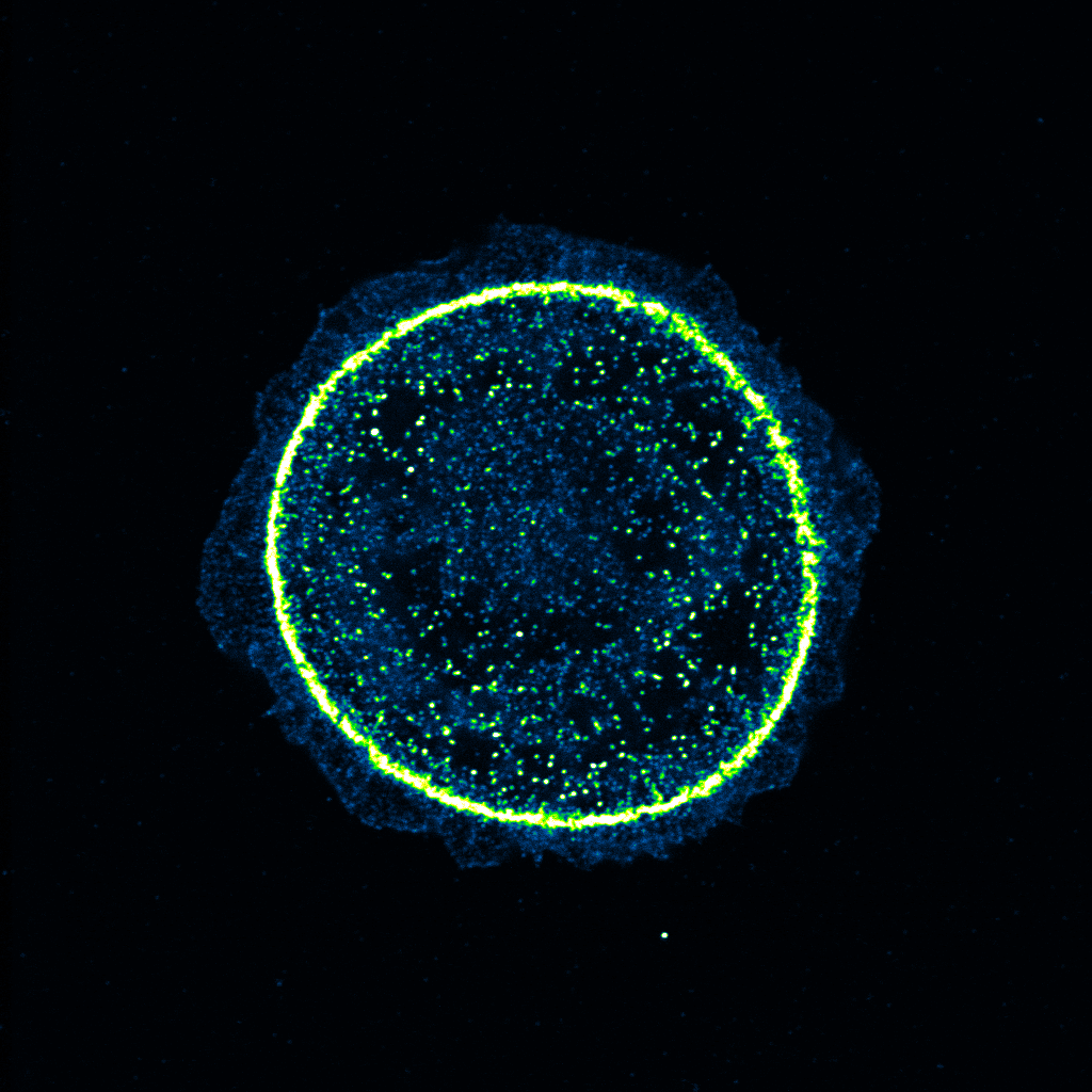
The ECM mainly consists of a network of protein fibres such as collagen and other filamentous extracellular proteins. In order to adhere to this meshwork, nearly every human cell possesses surface proteins termed integrins. Integrins operate like molecular carabiners that lock the cells to the network of collagen fibres or other ECM proteins and thus provide a strong focal point of attachment for the cell. However, cells in our body do not always stay in place, but sometimes need to migrate over long distances to their final destination – just think of immune cells that have to travel from the lymph node to a skin wound. As a solution, nature provided integrins with special features.
Like a sailor in the ship’s rigging
Integrins are peculiar, because they can be repeatedly folded over and extended: When folded over, integrins cannot connect to the ECM, as the carabiner is buried. Upon extension of the integrin, the carabiner is exposed and can lock the cell to the ECM. Interestingly, cells can extend integrins at the front end of the cell, while they fold over their integrins at the rear and detach from the ECM at these positions. Cycles of integrin-mediated “gripping and let loose” allow cells to move in the protein meshwork of the extracellular matrix like a sailor climbs in the ship’s rigging. Integrins consist of two parts, the α- and the β-subunit. Both subunits traverse the cell membrane, so that a small part of the protein is inside the cell, but the larger portion, the actual carabiner, is outside of the cell. It has been known for some time that the extension of the integrin is initiated by the integrin β-subunit: In particular, the protein talin initially binds to the intracellular part of the β-subunit and triggers integrin extension and thus the activation of the carabiner.
Without phosphorylation the carabiner hook remains buried
Tanja Grimm and Nina Dierdorf, doctoral researchers at the Konstanz Research School Chemical Biology have discovered that the β-subunit is being marked for talin binding by a small chemical modification, a so-called phosphorylation. This phosphorylation works like a switch: Upon phosphorylation, talin can bind and the integrin is extended. If phosphorylation does not take place or if this position in the integrin is mutated, talin does not associate and the carabiner remains buried. Consequently, the cells lose their grip. Moreover, the doctoral researchers now showed for the first time that a single enzyme is responsible for reversing the phosphorylation of the integrin β-subunit: the protein phosphatase PPM1F. This enzyme can remove the phosphorylation and thus trigger the integrins to fold over. The PPM1F-regulated “phosphorylation switch” in the integrin seems to be essential, because in the absence of PPM1F, embryonic development, when different cell types have to arrange themselves into functioning tissues, terminates prematurely. Isolated cells, in which the PPM1F gene is disrupted, show enhanced attachment to the extracellular matrix and can hardly move, as they are unable to release integrin-based matrix contacts.
Does less cell adhesion allow uncontrolled migration?
The researchers now hope that this knowledge can be used in the future to specifically control PPM1F activity and thus the functionality of integrins. In some tumour cells, this phosphatase appears to be particularly abundant, and the resulting reduced adhesion of such tumour cells could be one of the reasons, why they are able to leave the primary tumour and form metastases at distant body sites.
“In the next step, we want to learn how to manipulate the phosphorylation switch and thus the function of integrins," says Tanja Grimm, first author of the study. “We might be able to specifically influence integrin-dependent processes in our body, from immune cell movement to tumour metastasis. With these novel findings we might help our cells to firmly stay in touch with their surrounding and prevent them straying away for the worse”.
Key facts:
- Original publication: Grimm, T.M., Dierdorf, N.I., Betz, K., Paone, C., Hauck, C.R. (2020): PPM1F controls integrin activity via a conserved phospho-switch, Journal of Cell Biology (2020) 219 (12): e202001057; published online on 29 November 2020. doi: https://doi.org/10.1083/jcb.202001057
- The team led by Professor Christof Hauck, cell biologist at the Department of Biology, University of Konstanz, discovered new details about the regulation of cell adhesion and cell migration
- The phosphatase PPM1F is key for releasing integrin-based contacts between human cells and the extracellular matrix
- Research was conducted in the context of the Collaborative Research Centre (SFB 969) “Chemical and Biological Principles of Cellular Proteostasis”, which is funded by the German Research Foundation (DFG)
- Tanja Grimm and Nina Dierdorf, the lead authors of the study, received scholarships of the Konstanz Research School Chemical Biology (KoRS-CB).